
Beginner’s Guide to X Ray Fluorescence
What X Ray Fluorescence Is — and Why It Matters for Elemental Analysis
X-ray fluorescence (XRF) is a non-destructive analytical technique used to determine the elemental composition of materials — from metals and soils to liquids and powders — without destroying the sample.
If you’re looking for a quick answer, here’s what you need to know:
- What it does: Identifies and quantifies elements in a sample by measuring characteristic X-rays emitted when the material is excited by a primary X-ray source
- What it can detect: Elements from beryllium (Z=4) to uranium (Z=92), with practical routine analysis from sodium (Z=11) upward
- Detection limits: Typically a few parts per million (ppm); high-performance systems reach sub-ppm levels
- Sample types: Solids, powders, liquids — generally greater than 1 gram
- Key advantage: Fast, accurate, multi-element analysis with little or no sample preparation
- Main instrument types: Energy Dispersive XRF (EDXRF) and Wavelength Dispersive XRF (WDXRF)
For lab supervisors and technical managers who need reliable trace-element data fast, XRF is one of the most cost-effective and versatile tools available. It’s used across aerospace, pharmaceuticals, environmental testing, mining, and more.
This guide covers everything you need — the physics, instrument types, applications, sample prep, safety, and limitations — in plain language.
Understanding the Physics of X Ray Fluorescence
At its heart, X-ray fluorescence is a story of cosmic musical chairs played at the atomic level. To understand how we can identify a material simply by shining light on it, we have to look at how atoms interact with high-energy radiation.
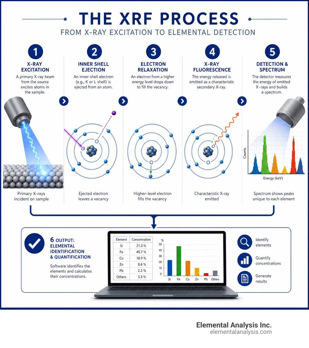
The process begins with a primary radiation source. Inside an XRF instrument, an X-ray tube (often utilizing targets made of rhodium, tungsten, molybdenum, or chromium) generates high-energy primary X-rays. When these energetic photons strike the target sample, they don’t just bounce off; they collide with the atoms inside the material, triggering a cascade of atomic events that results in the emission of secondary X-rays.
These secondary emissions are what we call characteristic radiation. They are unique to each individual element on the periodic table. The foundational physics behind this phenomenon were mapped out by Henry Moseley in the early 20th century. Moseley’s law established a direct, linear relationship between the square root of the frequency of the emitted X-ray and the atomic number of the element. In simple terms: heavier elements emit higher-energy, shorter-wavelength X-rays, while lighter elements emit lower-energy, longer-wavelength X-rays. This law is the mathematical bedrock that allows modern software to translate raw XRF spectra into highly accurate elemental percentages.
For a deeper dive into the physics and practical setup of this technology, you can download our comprehensive Get Glowing with This Complete X-Ray Fluorescence PDF Guide.
The Atomic Mechanism of X Ray Fluorescence
To understand exactly how these characteristic X-rays are born, we have to zoom in on the structure of the atom itself.
Atoms are structured like miniature solar systems, where a dense nucleus is orbited by electrons arranged in specific, nested orbital shells (designated as K, L, M, and so on, moving outward from the nucleus). The electrons in the innermost K shell are held with the strongest binding energy.
- Electron Ionization: When a high-energy primary X-ray photon strikes an atom, it must possess energy greater than the binding energy of an inner-shell electron to interact. If it does, the photon transfers its energy to an inner-shell electron (typically from the K or L shell), ejecting it entirely from the atom. This process of electron ionization leaves the atom in an unstable, highly excited state with an empty “vacancy” in its inner shell.
- Energy Transition: Nature abhors a vacuum, and atoms abhor instability. To restore balance, an electron from a outer, higher-energy orbital shell (such as the L or M shell) immediately drops down to fill the vacancy.
- Fluorescent Emission: Because outer-shell electrons possess higher energy states than inner-shell electrons, the migrating electron must shed its excess energy to fit into its new, lower-energy home. It releases this exact energy difference in the form of a fluorescent photon.
Because the energy spacing between orbital shells is a fixed quantum property unique to each element, the emitted photon’s energy acts as an atomic fingerprint. If an electron drops from the L shell to the K shell, it produces a K-alpha (Kα) X-ray. If it drops from the M shell to the K shell, it produces a K-beta (Kβ) X-ray. By measuring these precise energies, we can confidently identify which elements are present in the sample.
EDXRF vs WDXRF: Comparing Spectrometer Types
Not all XRF instruments are built the same way. Depending on how the instrument sorts and measures the emitted secondary X-rays, spectrometers are divided into two primary categories: Energy Dispersive XRF (EDXRF) and Wavelength Dispersive XRF (WDXRF).
Choosing between these two technologies is a matter of balancing your budget, required energy resolution, and the complexity of your samples. If you are looking to purchase or upgrade your laboratory capabilities, check out our review of The Best X-Ray Fluorescence Equipment to Upgrade Your Analysis.
To help you visualize the differences, here is a breakdown of how these two spectrometer types compare:
| Feature | Energy Dispersive (EDXRF) | Wavelength Dispersive (WDXRF) |
|---|---|---|
| Primary Sorting Mechanism | Direct collection by semiconductor detector | Physical dispersion via analyzing crystals |
| Energy Resolution | Moderate (~120 eV to 150 eV) | High (~20 eV to 50 eV) |
| Detection Limits | ppm levels (typically 1–10 ppm) | sub-ppm to ppb levels |
| Element Range | Sodium (Z=11) to Uranium (Z=92) | Beryllium (Z=4) to Uranium (Z=92) |
| Instrument Size | Compact, benchtop, or handheld | Large, stationary laboratory units |
| Analysis Speed | Fast (seconds to minutes for full scan) | Extremely fast for dedicated elements (<30s) |
| Relative Cost | Lower capital and maintenance costs | High capital and specialized facility costs |
Energy Dispersive Spectrometry (EDXRF)
EDXRF spectrometers are the workhorses of rapid, on-site testing. Their defining characteristic is their simplicity: they do not use complex optical paths to sort X-rays before they reach the detector. Instead, the entire spectrum of secondary X-rays emitted from the sample is directed straight into a high-performance semiconductor detector.
Modern EDXRF systems rely heavily on silicon drift detectors (SDDs) or older silicon PIN detectors. When an X-ray photon strikes the detector, it generates a charge pulse proportional to the photon’s energy. High-speed pulse processing electronics then sort these incoming signals, assigning them to different energy channels to build a spectrum in real time.
The primary advantages of EDXRF are its compact size, ease of use, and lower cost. Because they have no moving optical parts, these systems can be miniaturized into handheld devices that weigh less than 2 kg. However, because the detector is hit by all emitted X-rays simultaneously, EDXRF has lower energy resolution and can struggle with overlapping spectral peaks in highly complex matrices.
Wavelength Dispersive Spectrometry (WDXRF)
When extreme precision, low detection limits, and light element analysis are non-negotiable, WDXRF is the gold standard. Unlike EDXRF, a WDXRF system physically separates the emitted secondary X-rays by wavelength before they ever reach the detector.
This physical separation is achieved using a goniometer setup and highly specialized analyzing crystals. The secondary X-rays are directed onto a crystal monochromator with a known lattice spacing (d-spacing). By rotating the crystal and detector at precise angles, the instrument utilizes Bragg’s law of diffraction to allow only one specific wavelength of X-ray to reach the detector at a time.
Because of this physical filtering, WDXRF achieves an incredibly narrow energy resolution (often around 24 eV compared to 145 eV for EDXRF). This virtually eliminates peak overlaps, allowing for highly accurate trace-element analysis. Additionally, WDXRF can utilize specialized synthetic multilayers to detect light elements down to beryllium (Z=4). The trade-offs are that WDXRF instruments are large, expensive, require stable laboratory environments, and require rigorous sample preparation to achieve their potential. For a comprehensive historical and technical reference on these optical setups, refer to the Encyclopedia of Materials Characterization.
Practical Applications and Sample Preparation

While XRF is celebrated as a “non-destructive” technique, that label comes with a small asterisk. While the X-rays themselves do not damage or alter the chemical structure of your sample, the physical state of the sample heavily influences the accuracy of your results.
To achieve high-precision quantitative results, laboratories must address matrix effects. Matrix effects occur when the elements surrounding your target element either absorb the emitted fluorescent X-rays or emit their own secondary radiation that excites the target element further (secondary enhancement).
To minimize these errors, samples must be prepared to be as physically homogeneous as possible. There are three common preparation paths:
- Loose Powders: The sample is ground to a fine powder and poured into a plastic cup sealed with a thin Mylar film. This is ideal for rapid, qualitative screening but suffers from particle-size matrix effects.
- Pressed Pellets: The ground sample is mixed with a binding agent (like wax or cellulose) and pressed under high pressure (typically 20 to 30 tons) into a flat, smooth disc. This is excellent for trace element analysis because the sample remains concentrated.
- Fused Beads: For the ultimate in matrix reduction, the sample is mixed with a borate chemical flux (such as lithium tetraborate) and melted in a platinum crucible at temperatures exceeding 1000°C. The molten mixture is poured into a mold to cool, creating a perfectly homogeneous glass bead. This completely eliminates particle size and mineralogical matrix effects, making it the preferred method for cement and mining industries.
For practical workflows on preparing these samples, check out The Ultimate List of Practical XRF Applications.
Industrial Uses of X Ray Fluorescence
Because of its speed and versatility, XRF is utilized across a vast array of global industries:
- Alloy Verification: In metal fabrication, aerospace, and petrochemical industries, positive material identification (PMI) is critical. Handheld XRF units allow operators to instantly verify alloy grades (such as identifying 316 vs 304 stainless steel) on the production floor to prevent catastrophic material failures.
- Scrap Metal Recycling: Scrap yards use portable XRF devices to rapidly sort incoming metals by composition, allowing them to separate high-value alloys from mixed scrap in seconds.
- Cement Production: Automated, high-precision WDXRF systems are integrated directly into cement plant quality control loops. They monitor the ratios of calcium, silicon, aluminum, and iron in raw meal and clinker every few minutes to ensure structural integrity.
- Environmental Screening: Environmental engineers use XRF to screen soils and sediments for heavy metal contamination (such as lead, arsenic, and cadmium) at remediation sites, getting instant answers without waiting days for laboratory wet chemistry.
- RoHS Compliance: Electronics manufacturers use micro-spot XRF to scan circuit boards and plastic components, verifying that hazardous substances like lead, mercury, and polybrominated biphenyls are below the strict regulatory thresholds mandated by the Restriction of Hazardous Substances (RoHS) directive.
Limitations and Complementary Techniques
While we love XRF for its incredible utility, every analytical tool has its boundaries.
First, XRF is inherently a surface analysis technique. Because low-energy secondary X-rays can only penetrate a few micrometers to millimeters of a material, your sample must be representative of the bulk. If you scan a gold-plated copper ring, XRF will read a disproportionately high gold content unless you grind the sample.
Second, XRF struggles with light elements. Elements lighter than sodium (Z=11) produce very low-energy X-rays that are easily absorbed by the air, the detector window, or even the sample itself. While high-end WDXRF systems under vacuum can reach down to beryllium (Z=4), quantification is difficult.
Third, XRF cannot distinguish isotopes or different oxidation states of the exact same element (for instance, it cannot tell Fe²⁺ from Fe³⁺).
When these limitations pose a challenge, we turn to complementary techniques. At Elemental Analysis Inc., located in Lexington, KY, we specialize in bridging these analytical gaps. As the first commercial laboratory to offer Proton-Induced X-ray Emission (PIXE), we provide a powerful alternative. Learn more about how this advanced technique works in The Ultimate Guide to Particle-Induced X-Ray Emission.
Frequently Asked Questions About XRF
What elements can XRF detect?
XRF can theoretically detect elements ranging from beryllium (Z=4) to uranium (Z=92). In practical, routine benchtop or handheld applications, detection typically starts at sodium (Z=11).
Detection limits for heavier elements are incredibly sensitive, often reaching down to a few parts per million (ppm). For example, a portable handheld analyzer can detect down to 2 ppm of lead in pure sand. High-performance, stationary laboratory WDXRF systems can push those limits even lower, achieving sub-ppm or hundreds of parts-per-billion (ppb) detection for specific elements.
Is XRF analysis completely non-destructive?
Yes, XRF is widely regarded as a non-destructive testing (NDT) method because the primary X-ray beam does not alter, burn, or chemically change the sample. Once the analysis is complete, the sample is left completely intact and can be stored, archived, or analyzed using other techniques.
However, as we discussed in the sample preparation section, achieving high-accuracy quantitative results often requires grinding rocks into powders or melting them into glass beads. In these cases, the testing is non-destructive to the chemistry, but the sample preparation is destructive to the physical form of the material.
What safety precautions are required for XRF?
Because XRF instruments contain miniature X-ray tubes, they emit ionizing radiation when active. While modern closed-beam benchtop systems are completely shielded and safe for office environments, open-beam handheld units require strict safety protocols and operator training.
We always emphasize the ALARA principle: keeping radiation exposure As Low As Reasonably Achievable. This is managed through three core pillars:
- Time: Minimize the time spent near an active primary beam.
- Distance: Stand back. Radiation levels drop off exponentially with distance.
- Shielding: Use the instrument’s built-in shielding and backscatter guards.
Never aim a handheld XRF analyzer at yourself or another person, and never hold a sample in your hand during testing. In Kentucky, portable and laboratory X-ray equipment must be registered and operated in compliance with local regulations. For a detailed roadmap on local compliance and licensing, you can review the Kentucky Cabinet for Health and Family Services Radiation Guide.
Conclusion
X-ray fluorescence remains one of the most powerful, rapid, and versatile analytical techniques available to modern industry. Whether you are verifying aerospace alloys on a factory floor or monitoring environmental contaminants in soil, XRF delivers the fast, multi-element data you need to make critical decisions.
At Elemental Analysis Inc., based in Lexington, Kentucky, we combine our deep expertise in classical analytical chemistry with cutting-edge testing capabilities to solve your toughest elemental puzzles. As a pioneer in non-destructive testing and trace element identification, we are committed to delivering fast turnaround times and highly competitive pricing.
Ready to elevate your material testing? Explore our specialized XRF Services or reach out to our team in Lexington today to find the perfect analytical solution for your project.